Macular Degeneration Treatments
Examination For Age Related Macular Degeneration
Treatment for Age Related macular Degeneration
A complete and comprehensive ophthalmic examination is important in the assessment of AMD.
Patients will receive vision testing, drops to dilate pupils, and a complete examination of the front and back of the eye. Pupillary dilation may create blurring, and therefore, it is often best if a driver accompanies the patient, although it is not absolutely required.
Testing
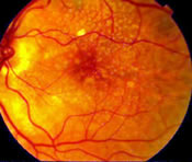
Patients with AMD may have several types of tests to assess the disease. Patients may undergo color photography of the macula and retina to document drusen, pigment changes, and other characteristics of AMD.
Fluorescein angiography (fluorescein angiography is performed by injecting sodium fluorescein dye into a peripheral vein with a small needle; this dye then goes through the body and eyes, as well as the macula to assess blood flow and to determine whether choroidal neovascularization is present) may be performed in the office to assess whether there is evidence of wet AMD. It is regarded as a safe test, but patients should expect some yellowish skin discoloration and orange urine. Most patients have no difficulty with this testing, although a low percentage of patients will experience some nausea. Any angiogram test, however, can be associated with allergic or even more severe reactions, and therefore, this test is reserved for patients in whom wet AMD is noted or suspected.
OCT imaging (OCT imaging is a non-invasive optical coherence tomogram examination of the macula; an OCT uses low energy laser to scan the macula and determine whether there is leakage beneath the macula or swelling within the macula, potentially signifying wet AMD) is a common non-invasive test used to assess the status of the macula. It can be used to determine whether there is evidence of choroidal neovascularization and wet AMD. It can also be used to assess response to treatment in patients with wet AMD. OCT is performed with no risk to the patient.
What the Doctor Sees
For patients with dry AMD, an ophthalmologist may note drusen accumulation within the macula, pigmentary changes within the macula, or advanced forms of dry AMD such as geographic atrophy. Patients with wet AMD are noted to have fluid in the macula, and hemorrhage or blood is commonly noted as well. There may be larger hemorrhages, and these are typically associated with sudden and severe vision loss. Other blister elevations (retinal pigment epithelial detachments) of fluid in the macula may or may not be symptomatic.
Prognosis For Age Related macular Degeneration
Most patients with AMD will retain good central vision and the ability to read in their lifetime. This is because 90% of patients have dry AMD. This is associated with a more favorable prognosis. Patients with wet AMD may suffer central vision loss. If it affects both eyes with associated vision loss, then the patient may progress to legal blindness, although AMD does not lead to total loss of vision. Thankfully, new treatments are improving the prognosis even for patients with advanced wet AMD.
Prevention and Treatment
Age Related macular Degeneration
Treatment of dry AMD is based on the Age Related Eye Disease Study (AREDS) which demonstrated that a specific formulation of anti-oxidant vitamins and minerals can reduce the risk of progression of dry AMD to more advanced stages and associated vision loss. Daily doses of vitamins in the AREDS study are:
500 mg Vitamin C
400 IU Vitamin E
15 mg Beta-carotene
80 mg Zink oxide
2 mg Copper oxide
It is important to check with your medical doctor before starting this AREDS supplement. In general, Vitamin E supplementation should not exceed 400 IU, and smokers should not be on any Beta-carotene supplementation due to an increase risk of lung cancer. There are AREDS formula vitamins available that do not have Beta-carotene that are appropriate for smokers.
Other recommendations noted above include:
- Stop smoking. Smoking has been associated with vision loss and more advanced forms of AMD.
- Eat a diet rich in colorful vegetables and fruits. These food groups have been associated with less advanced AMD and contain anti-oxidants which may slow progression of disease.
- Control blood pressure. High blood pressure is associated with more advanced vision loss and AMD.
- Consider sources of Omega 3 fatty acids. Omega 3 fatty acids found in cold water fish such as tuna and salmon, or those found in fish oil capsules are believed to be associated with less advanced AMD.
- Be physically active. Patients with AMD who were physically active several times a week were at reduced risk for advanced AMD and vision loss.
The prognosis for patients with wet AMD is improving although there are no cures for AMD. In 2006, Lucentis injection therapy was FDA approved for patients with wet AMD. This is the first treatment shown to improve vision in many patients with wet AMD. Monthly injections are typically performed by a retina specialist, and the procedure is relatively painless after eye drop anesthesia. Treatment may need to continue for up to 1 to 2 years depending on the nature of the wet AMD. Injections are typically performed in the office setting and carry only a low risk of associated problems such as hemorrhage or infection. Patients who receive injection therapy such as Lucentis will be asked to use topical antibiotics to reduce the risk of infection. Any patient who receives an injection and subsequently has increased pain or loss of vision should contact their doctor immediately.
Lucentis therapy works by blocking the growth of choroidal neovascularization and reducing leakage in the macula. Although approximately 70% of patients will maintain or improve vision, and 30% of patients have significant improvement in vision, there can still be vision loss despite Lucentis therapy. Again, Lucentis therapy may involve a series of eye injections, and in clinical trials at 2 years, patients had received 24 injections. Macugen therapy works similarly to slow the growth of choroidal neovascularization and to reduce leakage in the macula. Macugen was the first approved injection therapy for wet AMD.
Off-label Avastin injections are also being used to treat wet AMD. Avastin is a drug approved for use in cancer patients. Avastin is designed to shrink blood vessels and therefore shrinks certain cancer tumors after injection of the drug in a peripheral vein. Retina specialists have been performing direct injections of Avastin into the eye, similar to Lucentis and Macugen, to treat wet AMD. There is considerable clinical experience to demonstrate that Avastin may be helpful to patients with wet AMD, but scientific data are less rigorous compared to Lucentis or Macugen therapy. Any injection therapy, even when using only small doses in the eye, can potentially have side effects in the general body. Patients should discuss specific risks and benefits of treatments such as Avastin, Lucentis or Macugen with their eye specialist.
Photodynamic therapy (PDT) with verteporfin (Visudyne) utilizes an injection of a photosensitizing drug called verteporfin (Visudyne) and a non-thermal laser light to try and reduce leakage from certain types of choroidal neovascularization. It typically does not improve vision as a single treatment, however, it is being explored as an adjunctive agent to Lucentis therapy.
PDT is a timed office-based procedure. The Visudyne drug is infused into the vein over 10 minutes and then allowed to collect within choroidal neovascularization over another 5 minutes. The choroidal neovascularization is then exposed to a low energy laser light for about 90 seconds to activate the drug only within abnormal blood vessels in the macula. This produces a chemical reaction within these abnormal blood vessels to reduce blood flow and leakage.
Visudyne clears itself completely from the body over several days. The skin and eyes must be protected from sunlight with clothing and special sunglasses for 2 days after the treatment. No driving is allowed while wearing the sunglasses. Sometimes PDT is combined with other treatments such as Lucentis or with steroid injections to try and attack choroidal neovascularization from several different mechanisms of action.
Thermal laser photocoagulation may be used in certain cases of choroidal neovascularization where the abnormal blood vessels are not beneath the center of the macula. Thermal laser treatment attempts to heat and destroy choroidal neovascularization but also does affect overlying vision cells. Accordingly, this procedure is typically considered only when the blood vessels are far from the center of the macula. A major problem with thermal laser treatment is recurrent choroidal neovascularization which can develop in 50-60% of patients.
Low vision specialists help improve the quality of life for patients whose vision does not enable them to carry out daily activities. For example, AMD patients may have difficulty reading mail or seeing television clearly. Low vision rehabilitation is designed to use specific optical or lighting aids to try and improve specific vision tasks. Although low vision aids are no cure for AMD, they can be helpful in specific vision related tasks. Low vision specialists can also be a good resource for non-optical aids such as books on tape, large print reading materials, and writing guides.
Allen C. Ho, MD
2007.03.07